Dental Implant Failure — How to Prevent It, Spot It Early, and Treat It
When a dental implant no longer performs as it should — because it becomes loose, infected, or the surrounding bone and gum tissue decline — we call that implant failure. Failures happen for predictable reasons: incomplete osseointegration, peri‑implantitis (infection around the implant), mechanical overload from biting forces, or broader health issues that impair healing. This guide breaks down the causes, the early warning signs you can watch for, the clinical tests your dentist will use, and the treatment and prevention options so you can make informed choices. Catching a problem early often preserves bone and soft tissue and makes treatment simpler; waiting can mean implant removal and more extensive reconstruction. Read on for clear signals to watch, stepwise diagnostic checks (clinical exam and imaging), practical prevention steps, and how clinicians decide between non‑surgical care, surgical repair, or removal. If you’re worried about an implant or your risk factors, schedule a Free Dental Implant Consult with Dentist in Denton – Dentures & Dental Implants for a personalized assessment and next steps.
What Common Problems Cause Dental Implants to Fail?
Implant failure can occur early—during the bone‑healing phase—or later, months to years after placement. Early failures usually reflect failed osseointegration, where the implant never bonds solidly to the jawbone. Late failures most often stem from infection (peri‑implantitis), excessive mechanical forces, or gradual bone loss. Surgical factors (placement technique, implant position, initial stability) affect early outcomes, while patient factors—smoking, poor oral hygiene, uncontrolled diabetes—and bite forces influence long‑term success. Distinguishing early from late failure matters because options differ: early problems may be corrected or re‑implanted after healing, while late infections often need infection control and regenerative procedures. Below we outline the main mechanisms and how each undermines implant stability.
How Failed Osseointegration Causes Implant Problems
Failed osseointegration means the implant doesn’t form a direct, stable bond with the jawbone; instead a soft, fibrous layer prevents solid support. This can happen when the implant lacks primary stability at placement, the surgical site is traumatized, bone quality is poor, or the restoration is loaded too soon. Symptoms include persistent discomfort, a feeling that the tooth is loose, or lack of normal function during the expected healing window. Micromotion at the implant surface prevents new bone from forming and the implant may feel progressively looser. Clinicians reduce risk through careful site evaluation, securing strong primary stability, staged loading protocols, and by helping patients optimize controllable factors such as quitting smoking and improving blood‑sugar control.
Next is another common and preventable cause: infection‑driven peri‑implant disease.
What Peri‑Implantitis Is and Why It Matters
Peri‑implantitis is an inflammatory, bacteria‑driven condition that damages the soft tissue and underlying bone around an implant. It often begins as peri‑implant mucositis (reversible soft‑tissue inflammation) and progresses to bone loss if plaque and biofilm persist or the host response is dysregulated. Contributing mechanical factors include rough implant surfaces, residual cement, or excessive bite forces that trap plaque and cause microtrauma. Early clinical signs are bleeding on probing and deeper pockets around the implant; if left untreated, peri‑implantitis causes vertical and horizontal bone loss and can ultimately lead to implant mobility.
Microbial Dysbiosis in Peri-Implantitis: Pathogenesis and Immune Responses
ABSTRACT: Microbial dysbiosis in periodontitis and peri-implantitis: pathogenesis, immune responses, and therapeutic
Early recognition of peri‑implantitis is crucial: non‑surgical measures or timely surgical regeneration often save the implant, while delayed care frequently requires removal.
How Can You Tell an Implant Is Failing?
Detecting implant failure means watching for clinical, functional, and radiographic changes that deviate from normal healing and performance. Patients may notice persistent or worsening pain, swelling, or discharge. Dentists look for bleeding on probing, increasing pocket depths, and any mobility of the implant or its restoration. Changes in function—trouble chewing, a loose crown, or an altered bite—also warrant prompt review. Regular follow‑ups with periodontal charting and radiographs catch subtle bone and soft‑tissue changes before major problems develop. The sections below list early warning signals and explain how recession and bone loss appear clinically and on imaging.
Early Warning Signs of a Failing Implant
Early signs—often appearing weeks to months after placement—include pain that lasts beyond the expected healing period, persistent swelling, pus or any unusual drainage, and bleeding around the implant. Patients may describe sensitivity or a foreign‑body feeling. On exam a clinician may find redness, deeper probing depths, or suppuration when gentle pressure is applied. Even subtle mobility or micromotion is a red flag for poor osseointegration and calls for immediate assessment of loading and infection. Short‑term home care can include gentle antiseptic rinses and avoiding pressure on the area while you arrange a dental visit.
Quick clinical evaluation at this stage increases the chances of conservative salvage.
What Gum Recession and Bone Loss Look Like
Recession around an implant can expose the abutment or metal components, alter appearance, and create plaque‑trapping niches that raise infection risk. Clinically recession shows as a longer‑looking tooth or visible metal, and probing may reveal deeper pockets beside the recession. On radiographs early bone loss shows as crestal (vertical) reduction next to the implant and can progress to larger defects visible with three‑dimensional imaging. Spotting these changes early lets clinicians intervene with improved hygiene, targeted debridement, or regenerative procedures to protect implant stability.
Next we’ll cover the diagnostic tools dentists use to evaluate implants.
Which Strategies Help Prevent Implant Failure?
Prevention blends daily patient care, professional maintenance, and managing systemic or behavioral risks to support osseointegration and long‑term implant health.
Key elements are meticulous plaque control with appropriate interdental tools, routine professional cleanings to disrupt biofilm, and addressing modifiable risks such as smoking or uncontrolled diabetes.
Clinicians also reduce mechanical risks by using proper surgical technique, selecting implant sizes that match available bone, and planning the occlusion to prevent overload.
Below you’ll find practical hygiene steps, lifestyle recommendations, and a risk‑factor table that links common risks to targeted prevention tactics so patients know which actions matter most.
Removing biofilm around the crown, abutment, and gingival margin every day lowers the bacterial burden that causes mucositis and peri‑implantitis. Interdental brushes sized to the embrasure, soft‑bristled brushing, and adjunctive antimicrobial rinses help keep the area clean between professional visits.
Regular recall appointments let clinicians monitor probing depths, catch early bleeding on probing, and deliver focused debridement before bone loss progresses. These hygiene habits, combined with lifestyle changes, form a strong foundation for implant longevity.
How Good Oral Hygiene Lowers Failure Risk
Good oral hygiene controls the biofilm that drives inflammation and bone loss around implants. Daily use of an interdental brush sized to your implant restoration, gentle brushing (manual or powered), and adjunct antimicrobial rinses disrupt plaque at the crown‑abutment junction where bacteria gather. Your clinician will recommend specific tools and show techniques tailored to your prosthesis shape so hard‑to‑reach areas are cleaned effectively. Professional maintenance—often every three to six months depending on risk—removes hardened deposits patients can’t reach and checks soft‑tissue health. Consistent hygiene reduces mucositis and the chance it progresses to peri‑implantitis.
Lifestyle Changes That Protect Implants
Simple lifestyle changes make a big difference: quitting smoking, improving blood‑sugar control for diabetics, and addressing teeth‑grinding reduce complications and aid healing. Smoking lowers local circulation and immune response, impairing osseointegration and increasing infection risk — quitting or reducing tobacco use is one of the most impactful steps you can take. If you grind your teeth, a night guard and targeted occlusal adjustments lower excessive forces that damage peri‑implant bone. Good nutrition and medical management of systemic inflammation also support tissue repair. Together with daily hygiene and professional care, these changes strengthen long‑term outcomes.
Below is a concise table linking common risk factors to their effects on implants and practical prevention steps.
Risk Factor
Effect on Implant
Prevention / Management Strategies
Smoking
Reduces healing and raises infection risk
Smoking cessation programs; consider delaying elective implant surgery until abstinence improves outcomes
Poor glycemic control (Diabetes)
Slower healing and higher infection rates
Coordinate with medical providers to optimize blood sugar before and after surgery; close monitoring
Poor oral hygiene
Biofilm buildup leading to mucositis/peri‑implantitis
Daily interdental cleaning and professional maintenance every 3–6 months
Bruxism (teeth grinding)
Excessive load and micro‑movement at the implant
Night guard therapy, occlusal adjustments, and stress‑reduction strategies
Residual cement
Local irritation and plaque retention
Use screw‑retained restorations when appropriate or follow cementation best practices and remove excess carefully
This comparison shows how targeted actions reduce the specific ways each risk factor harms implants. Proactive management lowers complication rates and extends prosthetic life.
How Do Dentists Diagnose Implant Failure Early?
Diagnosis follows a stepwise approach that combines focused clinical testing with the right imaging to assess soft tissue, pocket depth, mobility, and bone levels. The initial step is a targeted exam—probing depths, checking for bleeding on probing, and testing mobility—followed by periapical radiographs to assess crestal bone. When two‑dimensional films don’t answer questions about bone volume or three‑dimensional defects, a CBCT (3D CT) scan gives detailed volumetric data needed for regenerative planning or deciding on removal. Routine monitoring and timely escalation to advanced imaging when clinical signs change are key to early detection. The sections below describe the clinical exam components and compare periapical X‑rays with CBCT for implant assessment.
Which Clinical Exams Reveal Implant Issues?
Clinical assessment includes a systematic check of peri‑implant soft tissues, probing depths, bleeding on probing, and mobility testing to identify instability. Measuring pocket depths with a periodontal probe and noting bleeding or suppuration quantifies inflammation and infection severity. Mobility testing differentiates between loose prosthetic parts (screws or crowns) and true fixture mobility, which signals lost osseointegration. Occlusal analysis—using shimstock or articulating paper—finds premature contacts or heavy forces that cause micro‑motion. Abnormal findings prompt radiographic evaluation and, when needed, specialist imaging or surgical consultation.
When X‑rays and CBCT Help
Periapical and bitewing radiographs are cost‑effective, low‑radiation tools for routine monitoring of crestal bone and the implant‑abutment interface, allowing side‑by‑side comparisons over time. If plain films are inconclusive or three‑dimensional detail is required—such as buccal or lingual bone defects, cortical plate integrity, or complex defect shapes—a CBCT scan provides the volumetric detail critical for surgical planning. CBCT is especially useful when deciding whether bone grafting or staged reconstruction is needed. Clinicians balance radiation exposure and cost against diagnostic benefit, using CBCT selectively to guide optimal treatment decisions.
What Treatment Options Exist for Failed Implants?
Treatment ranges from conservative non‑surgical care to surgical removal and staged reconstruction that rebuilds bone and allows future implants. The appropriate path depends on the failure stage—failed osseointegration versus advanced peri‑implantitis with significant bone loss—and patient factors like overall health, bone volume, and prosthetic needs. Non‑surgical care includes mechanical debridement, antiseptic therapy, and targeted antibiotics when indicated. Surgical options range from open debridement with regenerative grafting to implant removal with socket preservation and delayed re‑implantation. Patients should understand recovery timelines, invasiveness, and likely outcomes so they can take part in shared decision‑making.
When Non‑Surgical Care Is Suitable
Non‑surgical management fits early disease—peri‑implant mucositis or shallow peri‑implantitis with minimal bone loss and a stable implant. Conservative care includes careful mechanical debridement (using titanium or plastic instruments to avoid surface damage), antiseptic irrigation, and selective antimicrobial therapy when infection signs warrant it. Success depends on excellent patient plaque control and close follow‑up over weeks to months to confirm inflammation has resolved and probing depths stabilized. If there’s no improvement in pockets or radiographic bone levels, escalation to surgical regeneration or removal may be necessary.
Surgical Treatments for Implant Failure
Surgical options include open debridement with implant surface detoxification, regenerative surgery using bone grafts and membranes to rebuild defects, and implant removal followed by socket preservation and staged re‑implantation when appropriate. During regenerative procedures, clinicians remove granulation tissue, decontaminate the site, and place graft material with a barrier membrane to encourage new bone formation. When an implant cannot be saved because of severe loss or recurrent infection, careful removal aims to preserve as much native bone as possible and sets the stage for reconstruction or alternatives. Recovery focuses on soft‑tissue healing with radiographic reassessment before any re‑implantation is attempted.
Below is a quick comparison of treatment choices, how invasive they are, typical recovery times, and expected results to help you weigh options.
Treatment Option
Typical Indication / Invasiveness
Recovery Time
Typical Outcome
Non-surgical debridement
Early peri‑implantitis / Low invasiveness
Days–weeks
Often resolves inflammation with good patient compliance
Surgical regenerative therapy
Moderate–advanced bone defects / Moderate invasiveness
Weeks–months
Can restore bone and retain the implant in many cases
Implant removal and socket preservation
Severe bone loss or recurrent infection / High invasiveness
Weeks–months
Removes infection source; prepares site for staged reconstruction
Staged re-implantation after grafting
After successful grafting with adequate bone / High invasiveness
Months (healing)
High likelihood of success when bone is satisfactorily restored
How Bone Grafting Helps When Replacing an Implant
Bone grafting rebuilds deficient bone shape and volume, creating a biologic scaffold that supports later implant placement and reliable osseointegration. Options include autograft (your own bone), allograft, and xenograft; each has trade‑offs in regenerative potential, availability, and healing time. After grafting, a consolidation period of several months is usually needed for new bone to form and mature before attempting re‑implantation. Successful grafting improves the predictability of future implants by restoring ridge contours and providing stable bone where an implant can be positioned securely. Knowing graft choices and expected healing milestones helps patients understand the staged nature of reconstruction.
What Is Peri‑Implantitis and How Is It Managed?
Peri‑implantitis is an inflammatory disease that affects the tissue around an implant and causes progressive loss of supporting bone. Unlike peri‑implant mucositis (soft‑tissue inflammation only), peri‑implantitis involves bone destruction that becomes irreversible without prompt treatment. It arises from a mix of biofilm accumulation, host susceptibility, and local mechanical factors that encourage plaque retention and tissue breakdown. Treatment follows a stepped approach: mechanical and antiseptic measures for mild disease, localized antibiotics when appropriate, and surgical regeneration for moderate to advanced defects. The sections below explain common causes and summarize the most effective therapies.
Causes and Symptoms of Peri‑Implantitis
Peri‑implantitis is primarily driven by an infectious biofilm on implant surfaces together with an inflammatory host response that breaks down tissue and bone. Contributing elements include poor plaque control, residual cement, ill‑shaped restorations that trap debris, smoking, and systemic conditions that affect immunity. Symptoms include bleeding on probing, pus, deeper pockets, swelling, and discomfort; radiographs typically show crestal bone loss next to the implant. Disease progression depends on the balance between the biofilm challenge and host defenses, so prevention emphasizes plaque control and removing local mechanical contributors.
Most Effective Treatments for Peri‑Implantitis
Treatment depends on how advanced the disease is. Early peri‑implantitis can often be managed with mechanical debridement and improved hygiene. More advanced bone loss generally requires open surgical debridement and guided bone regeneration, sometimes combined with implant surface modification. Adjunctive options—localized antiseptics, laser therapy, or systemic antibiotics—are used selectively based on culture results, clinical severity, and individual patient factors. Surgical regeneration aims to rebuild lost bone and re‑establish a stable peri‑implant environment, enabling re‑osseointegration in many cases. Long‑term maintenance and frequent monitoring after treatment are essential to prevent recurrence.
How Long Do Implants Last and What Affects Their Lifespan?
When cases are well planned and maintained, dental implants have excellent long‑term survival—commonly reported between the low 90s and high 90s percent over five to ten years. Longevity depends on modifiable behaviors (oral hygiene, smoking), and fixed factors like bone quality and implant location. Regular maintenance visits, prompt care for peri‑implant inflammation, and attention to occlusal forces all boost the chances of long‑term success. The sections below put success rates in context and offer a table that links key longevity factors to their typical impact so patients can prioritize what matters most.
Typical Success Rates for Dental Implants
Under ideal conditions, implant success rates generally fall between about 90% and 98% over five to ten years, with variation by anatomical site, patient health, and prosthetic design. Success means the implant is stable, free of pain or infection, esthetically acceptable, and maintains bone levels; patient satisfaction with chewing and comfort is also a core measure. Cases with compromised bone, uncontrolled systemic disease, or poor maintenance show lower survival rates. These metrics set realistic expectations and highlight why prevention and risk management are so important.
Risk Factors That Shorten Implant Lifespan
Several factors shorten implant lifespan, and many are modifiable: smoking raises infection and failure risk; poorly controlled diabetes impairs healing; inadequate plaque control leads to chronic inflammation; and severe bruxism subjects implants to damaging overload. Local issues such as insufficient bone volume, thin gum tissue, or residual cement around restorations also increase complication risk. Clinicians can address local issues with improved technique, proper implant selection, and prosthetic design, while patients can reduce systemic risks through smoking cessation and medical optimization. Focusing on modifiable risks offers the most immediate benefit for long‑term success.
Below is a concise comparison of factors, their impact on longevity, and typical evidence or effect size to help prioritize risk management.
Factor
Impact on Longevity
Evidence / Typical Effect
Smoking
Significantly increases failure risk
Strong evidence; quitting improves outcomes
Oral hygiene/maintenance
Directly lowers peri‑implant disease
High impact; regular recall reduces failure rates
Diabetes (uncontrolled)
Slows healing and raises complication rates
Moderate–high impact; medical control reduces risk
Bone quality/volume
Shapes primary stability and long‑term support
High impact; bone grafting mitigates deficits
Occlusal overload/bruxism
Leads to mechanical failure and micro‑motion
Moderate impact; night guards lower risk
Why Choose Dentist in Denton for Implant Prevention and Treatment?
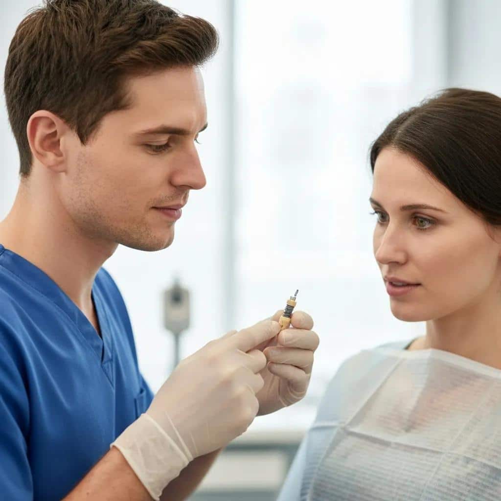
Dentist in Denton – Dentures & Dental Implants delivers education‑focused care in a single, convenient clinic, offering everything from diagnosis to advanced surgical correction and restoration in a calm, patient‑centered environment. We emphasize clear education so you understand your diagnosis, your options, and practical prevention steps — empowering you to take an active role in protecting your implants. If you have concerns, the practice offers a Free Dental Implant Consult that includes a focused assessment and a discussion of likely diagnostic steps and treatment paths. Our coordinated team and experienced clinicians make evaluation and treatment planning efficient for Denton‑area patients seeking timely care.
How Our Free Dental Implant Consult Helps
The Free Dental Implant Consult is a no‑cost first visit that focuses on listening to your concerns, reviewing your medical and dental history, and performing a targeted clinical check for obvious implant issues. During the consult we assess soft tissues, test for mobility and occlusal problems, and advise whether radiographs or 3D imaging are needed. That helps you prioritize next steps without upfront expense. We’ll outline potential treatments—from conservative care to surgical reconstruction—explain timelines and likely outcomes, and help you decide on the best path. Scheduling the Free Dental Implant Consult turns concern into a clear diagnostic plan and timely advice.
Meet Dr. Mike Pham and Dr. Vo
Dr. Mike Pham and Dr. Vo lead implant evaluation and treatment planning at Dentist in Denton – Dentures & Dental Implants, working within our patient‑first, education‑based model. Both doctors take part in comprehensive consultations to review findings, explain diagnostic options, and help patients weigh non‑surgical versus surgical treatments when implant issues arise. Their collaborative approach provides continuity from diagnosis through any treatment and ongoing maintenance, simplifying care for patients. To meet the team and start a tailored plan, request the Free Dental Implant Consult or call us at +16028340381.
Dentist in Denton offers a Free Dental Implant Consult to begin the diagnostic process.
The consult helps determine whether routine monitoring, imaging, or immediate treatment is needed.
Dr. Mike Pham and Dr. Vo collaborate to create clear, patient‑centered treatment plans.
These points show how our practice combines education, convenience, and a clear first step to care — and how acting early often prevents more invasive procedures later.
Early detection matters: Notice pain, swelling, bleeding, or mobility and seek evaluation promptly.
Prevention is proactive: Daily implant‑specific hygiene, smoking cessation, and maintenance visits reduce risk.
Timely diagnostics guide outcomes: Clinical exams plus periapical radiographs or CBCT inform the right treatment.
These final steps summarize how you can protect your implants and why the Free Dental Implant Consult at Dentist in Denton is a sensible next move if you have concerns about implant health.
Frequently Asked Questions
What are the long-term effects of dental implant failure on oral health?
When an implant fails, surrounding bone can resorb and gum tissue may recede, which can change bite relationships and affect nearby teeth. Functional issues—difficulty chewing or speaking—and esthetic concerns often follow, and additional procedures may be required to restore function and appearance. Early intervention reduces these long‑term impacts.
How can I prepare for a dental implant consultation?
Bring a list of current medications and medical conditions, any previous dental records or imaging, and your questions or concerns about the implant or the procedure. Being prepared helps the dentist evaluate your situation efficiently and provide tailored recommendations during the consult.
What lifestyle changes can enhance the success of dental implants?
Key changes include quitting smoking, eating a balanced diet that supports healing, and managing stress to reduce habits like teeth grinding. Smoking in particular impairs healing and raises infection risk. Addressing these areas supports tissue repair and long‑term implant success.
Are there specific foods to avoid after getting dental implants?
Avoid hard, crunchy, or sticky foods that place stress on the healing site (nuts, hard candies, chewy sweets, and tough meats). Very hot or spicy foods may also irritate healing tissues. Choose soft, easy‑to‑chew options—yogurt, mashed vegetables, smoothies—while you heal.
How often should I have follow-up appointments after implant placement?
Follow‑up visits are typically recommended every three to six months during the first year so your dentist can monitor healing, check implant stability, and spot early signs of complications. After the first year, recall frequency is adjusted based on your individual risk and oral health.
What should I do if I experience pain after dental implant surgery?
Contact your dentist right away if pain is severe or gets worse instead of improving. Some discomfort is normal after surgery, but worsening pain, swelling, or drainage can indicate infection or another complication that needs prompt evaluation and possible treatment.
Can dental implants fail years after placement?
Yes. Although long‑term success rates are high, implants can fail years later due to peri‑implantitis, mechanical overload, or systemic health changes. Ongoing maintenance, good hygiene, and managing health risks are essential to prevent late failures.
Conclusion
Knowing what causes implant failure and how to prevent it puts you in control of your oral health. Recognize early warning signs, keep up implant‑specific hygiene, and attend regular maintenance visits to maximize implant longevity. If you have concerns, schedule a Free Dental Implant Consult with our experienced team — we’ll review your situation, outline options, and help you protect your dental investment.