From Infection to Nerve Damage: Understanding Risks Associated With Dental Implants and How to Protect Yourself
Dental implants are titanium or ceramic fixtures placed into the jawbone to replace missing teeth, restoring chewing function and appearance while integrating with bone through osseointegration. Although dental implants have high success rates, two of the most concerning complications for patients are infection (including peri-implantitis) and nerve injury, which can cause persistent numbness or altered sensation. This article explains the mechanisms behind common implant risks, how to recognize early warning signs, and the diagnostic and treatment pathways clinicians use to protect oral function and sensation. You will learn the typical timeline for complications, how advanced imaging and surgical planning reduce nerve-related risks, and practical prevention steps to maximize long-term success with dental implants. The following sections cover the most common risks and a summary table, early infection signs and peri-implantitis, identification and management of nerve injuries, causes and early detection of implant failure, other less frequent complications, prevention strategies (including imaging and hygiene), when to seek professional help in Mansfield, TX , and frequently asked questions patients commonly ask.
What Are the Most Common Risks and Complications of Dental Implants?
Dental implants carry several distinct risks that range from early surgical problems to long-term biologic and mechanical complications. The primary complications include infection (surgical-site infections, peri-implant mucositis, peri-implantitis), nerve injury (inferior alveolar nerve, mental nerve, lingual nerve), implant failure from failed osseointegration or mechanical overloading, sinus perforation in the posterior maxilla, progressive bone loss, and rare allergic or hypersensitivity reactions to implant materials. Understanding the timing and typical presentation of each complication helps patients and clinicians prioritize prevention and early intervention to preserve the implant and oral health. The table below summarizes major complications, their common signs, likely timing after surgery, typical causes, and initial responses patients and clinicians should take when a problem is suspected. Reviewing these attributes prepares readers to recognize issues early and discuss targeted diagnostic plans with their provider.
The following table summarizes each major complication in a concise format to help with quick reference and triage.
Complication
Typical Signs
Initial Response
Infection / Peri-implantitis
Redness, swelling, bleeding on probing, pus, bad taste/odor
Seek clinical exam; antimicrobial rinses; possible antibiotics and debridement
Nerve Injury
Numbness, tingling, altered taste, sharp electric pain
Immediate clinical assessment; document sensory deficits; imaging and specialist referral
Implant Failure (osseointegration)
Persistent mobility, pain on biting, radiographic loosening
Avoid chewing on side; prompt evaluation and possible removal or revision
Sinus Perforation
Nasal discharge, sinus pressure, altered sinus symptoms after maxillary implant
ENT/dental evaluation; imaging; possible sinus repair or grafting
This summary clarifies how early recognition and proper initial responses differ across complications. Recognizing these patterns makes it easier to escalate care promptly and reduces the chance of irreversible damage, such as permanent nerve deficits or extensive bone loss that complicates later reconstruction.
Which Infections Can Occur After Dental Implant Surgery?
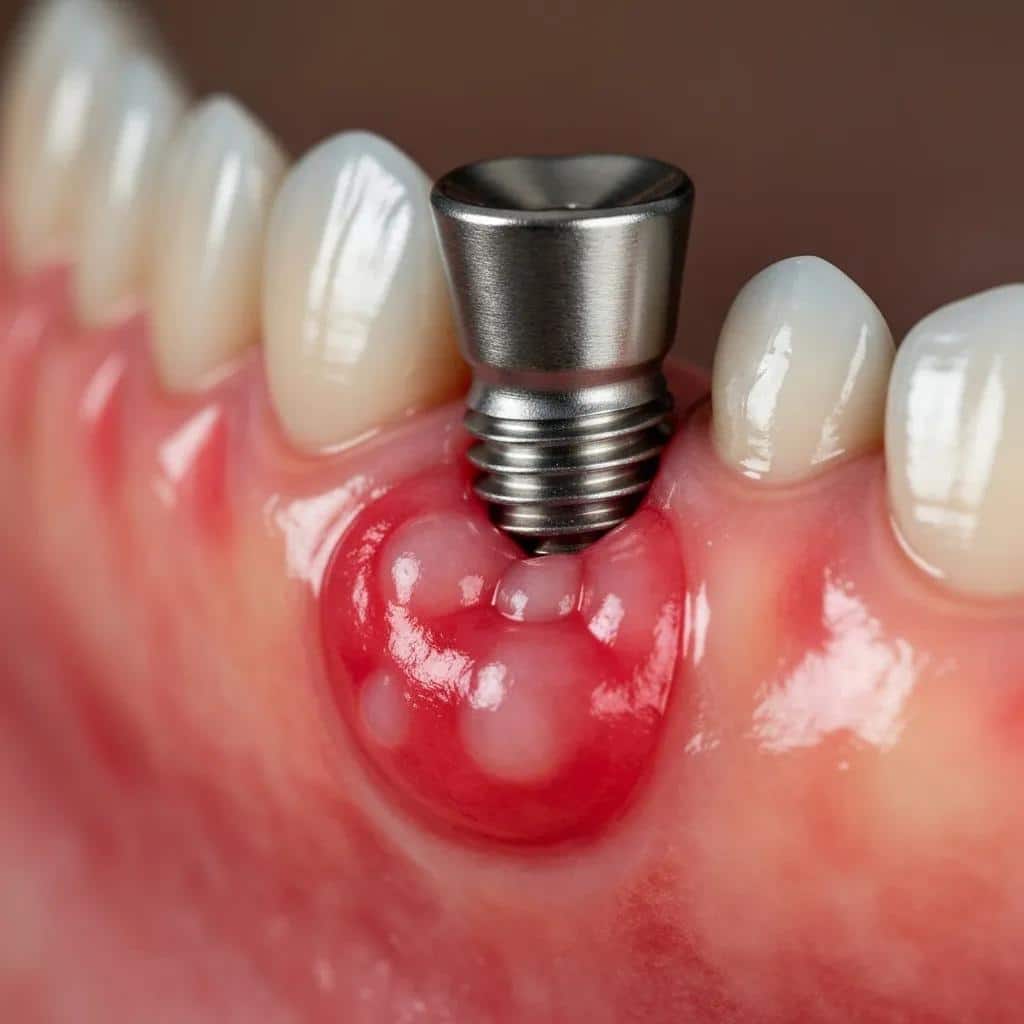
Infections after implant surgery range from superficial surgical-site infections to inflammatory conditions of the peri-implant tissues such as peri-implant mucositis and peri-implantitis. Peri-implant mucositis is an early, reversible inflammation of the soft tissues around an implant, typically caused by bacterial biofilm accumulation and poor oral hygiene, while peri-implantitis extends to bone loss and can lead to implant instability. Common causative organisms mirror periodontal pathogens—anaerobic gram-negative bacteria—and risk factors include smoking, uncontrolled diabetes, inadequate plaque control, and residual cement around implant crowns. Typical onset can be within days for acute surgical infections or months to years for peri-implantitis as plaque accumulates; early detection often relies on routine probing, radiographs, and clinical vigilance.
Patients benefit from a timeline example: immediate postoperative redness with mild pain that resolves within a week is often normal, while persistent or worsening pain, drainage, or increasing probing depths after two to four weeks should prompt re-evaluation. Early management emphasizes local debridement, improved hygiene, antiseptic rinses, and short courses of systemic antibiotics when indicated; persistent bone loss may require surgical intervention and regenerative approaches. Identifying the infection stage early improves the likelihood of resolving inflammation without implant loss and sets the stage for targeted preventive measures discussed later.
How Does Nerve Damage Happen With Dental Implants?
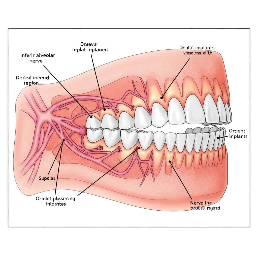
Nerve damage related to dental implants typically occurs through direct trauma, compression by implants or bone grafting materials, traumatic local anesthetic injections, formation of postoperative hematoma, or inadequate pre-surgical planning that places implants too close to neural structures. The inferior alveolar nerve runs within the mandibular canal and supplies sensation to the lower lip, chin and teeth; the mental nerve is a terminal branch providing lower lip and chin sensation, and the lingual nerve provides tongue sensation. When implant osteotomies or fixtures encroach on these pathways, sensory disturbances can result from neuropraxia (temporary conduction block) to more severe axonotmesis or transection, which carry worse prognoses.
Advanced imaging and careful surgical planning are critical because an anatomic millimeter can make the difference between a safe placement and a nerve injury. Using three-dimensional imaging and surgical guides reduces the likelihood of unintentional nerve contact, while conservative osteotomy depths and staged grafting strategies protect neural integrity. Timely recognition and documentation of altered sensation immediately after surgery allow clinicians to pursue early decompression, medication, and specialist referral options, improving the chance of meaningful recovery.
Inferior Alveolar Nerve Injury in Dental Implants: Causes, Symptoms, Diagnosis, and Treatment
The purpose of present article was to review aetiological factors, mechanism, clinical symptoms, and diagnostic methods as well as to create treatment guidelines for the management of inferior alveolar nerve injury during dental implant placement.
Injury of the inferior alveolar nerve during implant placement: a literature review, G Juodzbalys, 2011
What Are the Early Signs and Symptoms of Dental Implant Infection?
Early recognition of infection around implants dramatically affects outcomes and the ability to preserve the implant. The most common early signs include redness and swelling of the gums around the implant, persistent or increasing pain beyond the expected postoperative window, bleeding or pus on probing, a foul taste or odor, and increased probing depths on clinical exam. These symptoms often progress from soft tissue inflammation (mucositis) to bone-involving peri-implantitis if untreated, so patients should monitor healing carefully and seek prompt evaluation for persistent abnormalities. Below is a concise featured-snippet–style list of early signs optimized for quick recognition and patient action.
Early signs and what they mean are listed here for quick assessment and to guide early self-care and timely clinician contact.
Redness and swelling: Inflamed gum tissue around the implant that worsens over days rather than improving.
Persistent pain: Increasing or unrelieved pain that continues beyond the normal 7–10 day postoperative period.
Bleeding on probing: Ongoing bleeding when gently probing near the implant indicates active inflammation.
Pus or drainage: Any visible pus or persistent drainage is a clear sign of infection requiring urgent assessment.
Bad taste or odor: Foul taste or halitosis localized to the implant site suggests bacterial involvement.
Increased mobility: Early looseness of the implant or prosthetic components may accompany advanced infection.
These early warning signs differentiate normal healing from pathologic processes and should prompt avoidance of self-manipulation, maintenance of gentle hygiene, and a clinical visit for assessment and possible intervention. A clear next step is clinical probing, radiographic review, and implementation of appropriate antimicrobial measures or referral if the problem is advanced; prompt action reduces the likelihood of bone loss and implant loss.
What Is Peri-Implantitis and How Does It Affect Implants?
Peri-implantitis is an inflammatory disease characterized by soft-tissue inflammation around the implant and progressive loss of supporting bone. It begins with plaque-induced mucositis that, if unaddressed, progresses to bone resorption detectable on radiographs and increased probing depths clinically. Diagnosis typically combines clinical signs—bleeding on probing, suppuration, increased pocket depths—with radiographic evidence of bone loss compared to prior baselines; absence of prior images can complicate assessment but rising probing depths and symptoms are still concerning.
Treatment spans non-surgical debridement and antiseptic therapies for early disease to regenerative or resective surgical approaches for established bone loss; adjunctive systemic or local antibiotics may be used selectively based on severity and microbial patterns. Left unchecked, peri-implantitis can create progressive bone defects that undermine implant stability and ultimately necessitate removal and complex reconstruction, so early surveillance and regular maintenance care are crucial to preserve long-term implant success.
How Can You Recognize Symptoms Like Swelling, Pain, and Pus?
Distinguishing normal post-surgical discomfort from signs of infection requires a timeline-based approach and specific symptom descriptors. Normal postoperative swelling and mild pain typically peak within 48–72 hours and then gradually improve; by one to two weeks most acute symptoms should subside with appropriate self-care. Red flags include worsening or new-onset swelling after initial improvement, increasing intensity of pain despite analgesics, persistent or growing pockets of pus or drainage, and systemic signs such as fever or malaise.
Patients can perform simple self-checks—comparing the treated side with the opposite side, noting whether pain is increasing rather than decreasing, and checking for persistent taste changes or drainage. If any red flags are present, avoid manipulating the area, maintain gentle oral hygiene with antiseptic rinses as advised, and seek prompt clinical evaluation. Early professional assessment often includes clinical probing, radiographs or 3D imaging, and targeted debridement; acting quickly can halt progression and reduce the need for surgical rescue.
How Can Nerve Damage From Dental Implants Be Identified and Treated?
Nerve injuries from dental implants require systematic clinical assessment and often multimodal management to maximize recovery and reduce permanent deficits. Identification begins with a thorough sensory exam that documents numbness, tingling, altered temperature perception, dysesthesia, or sharp electric-like pains in the distribution of the inferior alveolar, mental, or lingual nerves. Clinicians use objective tests—two-point discrimination, light touch, pinprick, and thermal testing—along with patient-reported symptom mapping to establish the injury’s extent and timeline. Imaging, especially three-dimensional scans, helps determine whether the implant or graft material encroaches on neural canals.
Treatment depends on injury severity and timing: immediate decompression or repositioning may be indicated if the implant is impinging on a nerve and intervention occurs early. Conservative management includes anti-inflammatory medications, short courses of steroids when appropriate, neuropathic pain agents, close observation, and referral to oral and maxillofacial specialists or neurosurgical colleagues for persistent deficits. Early referral and documentation improve prognostic clarity; the following table compares common nerve injuries and their symptom profiles to assist patient understanding and clinical triage.
The table below compares inferior alveolar, mental, and lingual nerve injuries by affected area and typical symptom profile to aid rapid identification and targeted assessment.
Nerve
Affected Area
Symptom Profile
Inferior alveolar nerve
Lower teeth, lower lip, chin
Numbness/tingling of lower lip/chin, altered tooth sensation, possible pain on mastication
Mental nerve
Lower lip and chin skin
Localized numbness or paresthesia of the lower lip and chin; speech and eating may feel altered
Lingual nerve
Anterior two-thirds of tongue
Altered taste, tongue numbness, burning or dysesthesia during eating and speech
This comparison clarifies how symptom location maps to anatomy and helps patients describe their experience accurately to clinicians. Accurate symptom mapping often influences urgency: sudden extensive numbness or progressive neuropathic pain typically triggers faster escalation to imaging and specialist consultation.
Systematic Review: Inferior Alveolar Nerve Injury After Dental Implants – Diagnosis and Treatment Outcomes
The purpose of this article is to systematically review diagnostic procedures and risk factors associated with inferior alveolar nerve injury following implant placement, to identify the time interval between inferior alveolar nerve injury and its diagnosis after surgical dental implant placement and compare between outcomes of early and delayed diagnosis and treatment given based on case series recorded throughout a period of 10 years.
Inferior alveolar nerve injuries following implant placement-importance of early diagnosis and treatment: a systematic review, G Juodzbalys, 2014
What Are the Symptoms of Inferior Alveolar Nerve Damage?
Inferior alveolar nerve injury most commonly presents with numbness or tingling of the lower lip and chin, altered or reduced sensation of lower teeth, and sometimes uncomfortable dysesthesia described as electric shocks or burning. Functionally, patients may notice drooling, difficulty sensing food on the lip, altered speech articulation, and reduced proprioceptive feedback when biting. Symptoms onset can be immediate after surgery—suggesting direct trauma—or delayed if compression from hematoma or swelling develops; the prognosis varies with injury type, with neuropraxia often resolving over weeks to months while axonotmesis or transection may cause longer-term deficits.
Self-assessment techniques, such as comparing sensation with the contralateral side and noting changes in taste or tongue function, help in early reporting to the dental team. Early documentation and prompt referral for imaging and specialist evaluation improve the chance of meaningful recovery, especially when decompression or surgical repair is possible within a short window after injury.
What Treatment Options Are Available for Nerve Injury?
Treatment options range from observation and medical therapy to surgical decompression or repair, depending on severity and timing. Conservative strategies include anti-inflammatory medications, short courses of corticosteroids to reduce edema when appropriate, neuropathic agents (such as certain antidepressants or anticonvulsants) to manage dysesthesia, and close follow-up with repeat sensory testing. When imaging reveals implant impingement, hematoma, or hardware-related compression, early surgical intervention—removal or repositioning of the implant and direct nerve decompression—can improve outcomes; microsurgical nerve repair may be indicated for transections.
Rehabilitation may include sensory retraining and multidisciplinary collaboration with oral surgery, neurology, or pain specialists for persistent neuropathic pain. Importantly, early recognition and timely escalation are associated with better prognosis, which is why preoperative planning and intraoperative caution are essential components of prevention and why patients should report any sensory abnormalities immediately.
What Causes Dental Implant Failure and How Can You Spot It Early?
Implant failure has multifactorial causes that include biological, mechanical, and patient-related factors; spotting early warning signs can allow salvage strategies that prevent complete loss. Biologic causes center on failed osseointegration and infection—if bone fails to integrate with the implant surface or peri-implantitis progresses, the fixture can become mobile. Mechanical failure occurs from overloading, poor prosthetic design, or component fracture, while host factors such as smoking, uncontrolled diabetes, osteoporosis, or medications that affect bone metabolism increase failure risk. Early recognition of failure centers on detecting mobility, persistent pain, and radiographic changes.
A numbered list below outlines the top causes and what early signs to watch for, helping patients and clinicians prioritize evaluation and corrective steps.
Failed osseointegration: Signs include persistent implant mobility and lack of functional stability within months of placement.
Infection-driven bone loss: Presents as chronic inflammation, bleeding, suppuration, and progressive radiographic bone defects.
Mechanical overloading or poor prosthetic fit: Detect with pain on biting, restoration loosening, or visible prosthetic misalignment.
Systemic and behavioral factors: Smoking, poor glycemic control, and certain medications reduce bone healing capacity and increase complication risk.
This framework enables targeted monitoring: patients experiencing any of these early signs should minimize function on the affected side and seek prompt clinical evaluation for radiographic assessment and possible mechanical or biological interventions. Early prosthetic adjustments or non-surgical infection control can sometimes restore stability without removing the implant.
What Are the Common Reasons for Implant Failure?
Common reasons for implant failure fall into biological categories such as failed osseointegration and peri-implantitis, mechanical problems like overload or prosthetic misfit, and host-related issues including smoking, uncontrolled diabetes, and medications that impair bone metabolism. Failed osseointegration often reflects inadequate primary stability, poor bone quality, or micromotion during healing, whereas peri-implantitis stems from long-term biofilm accumulation and host inflammatory responses. Mechanical failure involves excessive occlusal forces, bruxism, or improper distribution of chewing loads leading to screw loosening or fractured components.
Examples include a patient with poor bone density who experiences early mobility due to insufficient primary stability, or a case where excess cement around the crown fosters chronic inflammation that evolves into peri-implantitis. Preventive measures—achieving primary stability, appropriate prosthetic design, and rigorous hygiene—map directly to these causes and reduce the probability of failure when implemented consistently.
What Are the Warning Signs of Implant Rejection or Loosening?
Warning signs include persistent or increasing pain localized to the implant site, new looseness of the crown or implant fixture, shifting occlusion, and repeated episodes of local inflammation or discharge despite hygiene measures. Radiographs showing widening of the peri-implant radiolucency or progressive bone loss are objective indicators of instability. Immediate practical steps include avoiding chewing on the affected side, maintaining gentle hygiene, and seeking a clinical evaluation that includes radiographic imaging and possibly removal of prosthetic components to inspect implant mobility.
Rapid reporting and assessment allow clinicians to determine whether non-surgical therapy, prosthetic adjustment, or surgical revision is necessary. If mechanical factors predominate, prosthetic redesign or occlusal equilibration may salvage the implant; if biological factors dominate, debridement and regenerative approaches might be required to re-establish stable bone support.
What Other Complications Can Arise From Dental Implants?
Beyond infection and nerve injury, implants can be associated with sinus membrane perforation, progressive peri-implant bone loss requiring grafting, rare allergic or hypersensitivity reactions, and prosthetic complications such as abutment screw loosening or crown fracture. These complications vary in frequency and severity but can significantly affect outcomes when they occur, particularly in anatomically challenging regions or when host factors compromise healing. Understanding how these issues present and are managed helps patients evaluate risk and informs pre-surgical discussions about mitigation strategies.
The following subsections examine sinus-related complications and the impact of bone loss and allergic reactions, illustrating practical signs and corrective pathways to maintain implant longevity.
How Do Sinus Problems and Sinus Perforation Affect Implant Success?
Sinus perforation most commonly affects implants placed in the posterior maxilla when available bone height is limited and the sinus membrane is breached during osteotomy or implant placement. Consequences may include sinusitis, persistent nasal discharge that is unilateral, a feeling of sinus pressure, or oroantral communication in severe cases. Preventive strategies include preoperative assessment of sinus anatomy with three-dimensional imaging, staged sinus lift procedures when indicated, and careful surgical technique to maintain membrane integrity.
When sinus involvement is suspected, timely evaluation with imaging and possible ENT collaboration is important; minor perforations may heal or be managed intraoperatively, while larger defects or chronic sinusitis may require sinus membrane repair or alternative implant-site rehabilitation. Proper planning and using sinus lift or grafting techniques when bone height is insufficient reduce the risk of intraoperative perforation and improve implant survival in the posterior maxilla.
What Is the Impact of Bone Loss and Allergic Reactions on Implants?
Progressive bone loss undermines implant stability by reducing the osseous support that anchors the fixture; left unchecked, it leads to mobility and potential implant failure. Bone grafting and regenerative procedures can rebuild lost support in many cases, but success depends on infection control, systemic health, and careful surgical technique. Allergic reactions to implant materials are rare but can present as persistent inflammation or unexplained mucosal reactions; diagnostic approaches include patch testing and material alternatives such as zirconia implants for those with confirmed sensitivity.
Signs that bone loss is progressing include deepening probing depths, exposed implant threads, and radiographic evidence of marginal bone reduction. Early detection paired with targeted interventions—debridement, antimicrobial therapy, and regenerative grafting—offers the best chance to stabilize the site and preserve the implant.
How Can You Prevent Dental Implant Risks and Ensure Long-Term Success?
Preventing implant complications requires a layered approach that spans pre-surgical assessment, meticulous surgical technique, and rigorous post-operative maintenance. Key measures include comprehensive pre-surgical planning with three-dimensional imaging to map nerve anatomy and bone dimensions, use of surgical guides to translate planning precisely to the operative site, adherence to sterile technique and careful soft-tissue management during surgery, and a structured post-op maintenance protocol emphasizing oral hygiene, smoking cessation, and regular professional cleanings and exams. Each preventive action targets specific risks—CBCT imaging reduces nerve and sinus complications, surgical guides lower placement errors, and diligent hygiene reduces peri-implantitis risk—so combining these strategies produces synergistic protection.
Below is a practical prevention-reference table linking concrete preventive measures to the specific risks they mitigate and simple steps patients and clinicians can implement.
Preventive Measure
What it Protects Against
Practical Steps
CBCT / 3D imaging
Nerve injury, sinus perforation
Request 3D scans for implant planning and identify nerve/sinus position
Surgical guides
Misplacement / nerve encroachment
Use guided osteotomies translated from the digital plan into surgery
Rigorous oral hygiene
Peri-implant mucositis / peri-implantitis
Daily brushing, interdental cleaning, antiseptic rinses, professional cleanings
Smoking cessation & systemic control
Osseointegration failure
Stop smoking before and after surgery; manage diabetes and systemic risks
This crosswalk clarifies how each practical step translates into measurable risk reduction. Patients should discuss these measures with their clinician before treatment to understand how each contributes to safer outcomes.
Free implants consultation : A brief note for patients weighing options — the practice Dentist In Mansfield- Dentures and Dental Implants (operating under the brand “Affordable Dentist Near Me”) offers a Free implants consultation as a non-pressured risk assessment resource to review imaging and discuss planning choices. This option can help patients confirm whether advanced imaging or surgical guides are recommended for their case before committing to surgery.
A structured prevention plan that combines these elements significantly reduces complication rates and supports predictable long-term success for dental implants, especially when patients adhere to maintenance and clinicians apply contemporary planning tools.
What Role Does Oral Hygiene Play in Preventing Infection?
Oral hygiene is foundational to preventing peri-implant mucositis and peri-implantitis because bacterial biofilm accumulation at the implant-abutment interface initiates inflammatory responses leading to tissue breakdown. Daily mechanical plaque control—soft-bristled brushing twice daily, interdental cleaning with brushes or floss designed for implants, and adjunctive antiseptic rinses—minimizes biofilm formation. Professional maintenance visits for implant-site prophylaxis and probing evaluation at regular intervals detect early inflammation before bone loss occurs.
Behavioral factors such as smoking and systemic conditions like poorly controlled diabetes increase susceptibility to infection and should be managed as part of the hygiene plan. Patients who prioritize consistent home care and attend routine professional cleanings substantially reduce their long-term risk of peri-implant disease and enhance the durability of their implants.
How Does Pre-Surgical Planning and Advanced Imaging Reduce Nerve Damage?
Pre-surgical planning with CBCT imaging maps bone volume, canal anatomy, and sinus position, allowing clinicians to select implant lengths and trajectories that maintain safe distances from neural structures. Digital planning software enables virtual implant positioning that accounts for restorative goals while avoiding nerve canals, and the use of printed or milled surgical guides transfers those plans accurately to the surgical field. These steps reduce intraoperative guesswork and the risk of inadvertent nerve contact or sinus breach.
A short example: a CBCT can reveal a shallower-than-expected mandibular canal position, prompting a change to a shorter implant or a staged grafting approach rather than risking canal proximity. Asking your clinician about 3D imaging and surgical guides during preoperative discussions ensures that nerve-protection strategies are part of the proposed plan and supports safer surgical execution.
When Should You Seek Professional Help for Dental Implant Complications in Mansfield, TX?
Knowing when to seek urgent or prompt care prevents escalation of implant problems into emergencies and reduces the potential for permanent harm. Immediate dental attention is required for severe signs such as uncontrolled bleeding, swelling that impairs breathing or swallowing, sudden onset of extensive numbness indicating possible nerve injury, or signs of systemic infection like fever and malaise. Prompt, non-emergency care is advisable for persistent or worsening pain, drainage, mobility of the implant or crown, and ongoing bad taste or odor localized to the implant area. The short checklist below helps patients decide when to seek care and what to expect when they contact a local clinic.
This action-oriented checklist emphasizes that early clinical assessment—especially when local services offer low-friction initial evaluation—shortens the time to definitive care, reduces the risk of progression, and helps patients make informed decisions about intervention.
Seek immediate care if you have any of the following: Severe swelling that affects breathing or swallowing
Uncontrolled bleeding from the surgical area
Sudden, widespread numbness or paralysis in the lip, chin, or tongue
Seek prompt evaluation for: Persistent pain beyond the normal healing window
Visible pus, ongoing drainage, or a bad taste that does not resolve
New mobility of the implant or prosthetic components
Interim measures before care: avoid chewing on the affected side, maintain gentle hygiene, and avoid manipulating the area
If you are in Mansfield and experiencing these symptoms, a Free dental consult can help triage the issue and plan next steps. For patients “Trying to schedule a free consult”, the offering from Dentist In Mansfield- Dentures and Dental Implants (operating under the brand “Affordable Dentist Near Me”) is positioned to evaluate symptoms, review imaging such as CBCT if available, and outline immediate management or referral pathways. A free consult typically includes a focused clinical exam, symptom triage, and discussion of recommended diagnostic imaging or specialist referrals to clarify next steps and reduce anxiety.
This immediate-action guidance emphasizes that early clinical assessment—especially when local services offer low-friction initial evaluation—shortens the time to definitive care, reduces the risk of progression, and helps patients make informed decisions about intervention.
What Symptoms Require Immediate Dental Attention?
Emergency symptoms include acute airway compromise from swelling, heavy uncontrolled hemorrhage, and sudden onset of extensive numbness that may indicate major nerve involvement. Severe progressive infection with systemic signs—fever, increasing malaise, or rapidly expanding swelling—also requires urgent assessment and possible systemic therapy. For these scenarios, patients should not delay contacting emergency or urgent dental services and should avoid applying pressure or manipulating the area, which could worsen bleeding or spread infection.
Non-emergency but concerning signs include persistent drainage, increasing localized pain, or new prosthetic mobility; these warrant timely appointments within days so clinicians can perform imaging, debridement, or prosthetic adjustments. Local availability of imaging and a structured triage process improves patient outcomes by accelerating diagnostic clarity and treatment decisions.
How Can a Free Consultation Help Manage Implant Risks?
A Free New Patient Consultation or Free implants consultation functions as a low-barrier first step to assess symptoms, review existing imaging, and plan diagnostic testing such as CBCT scans when necessary. During a consult, the clinician documents symptom onset, performs targeted sensory testing and oral exam, evaluates prosthetic fit, and recommends immediate or staged interventions based on findings. For patients uncertain whether symptoms constitute an emergency or manageable complication, the consult helps triage care, provides an evidence-based plan, and outlines potential next steps including referrals for advanced imaging or specialist care.
This non-pressured consult reduces anxiety by clarifying the problem, estimating urgency, and presenting treatment options in plain language—making it easier for patients to decide on timely action. If you are “Trying to schedule a free consult”, using that appointment to have your symptoms assessed, imaging reviewed, and a clear plan documented is an effective first step toward resolving implant problems with local, coordinated care.
What Are the Most Frequently Asked Questions About Dental Implant Risks?
Patients commonly ask about timelines for recovery, the probability of permanent damage, and whether allergies to implant materials are likely. Clear answers help set expectations and guide decision-making; this FAQ section addresses those recurring questions succinctly and with practical advice. Each answer balances evidence-based perspective with patient-centered clarity, giving readers concise guidance to bring to clinical discussions.
How Long Do Side Effects Like Infection or Nerve Pain Last?
Minor post-operative side effects such as mild swelling and transient pain typically resolve within one to two weeks as healing progresses. Superficial surgical infections that respond to antibiotics and local care often improve over days to a week, while peri-implantitis may require months of treatment and ongoing maintenance to control. Nerve symptoms vary widely: neuropraxia may improve within weeks to months, whereas more severe nerve damage can persist or require surgical repair and prolonged rehabilitation; persistent deficits beyond three months should prompt specialist evaluation. Factors that lengthen recovery include delayed presentation, ongoing smoking, poorly controlled systemic conditions, and untreated infection, so early intervention and risk-factor management shorten recovery trajectories.
This timeline helps patients set realistic expectations and reinforces the importance of follow-up if symptoms do not follow typical post-operative resolution patterns.
Can Dental Implants Cause Permanent Damage or Allergic Reactions?
Permanent nerve damage is uncommon but possible if there is direct nerve transection or prolonged compression without timely management; early detection and intervention reduce this risk. Allergic or hypersensitivity reactions to implant materials are rare; when suspected due to unexplained chronic inflammation or dermatologic signs, clinicians may consider material testing or alternative materials such as zirconia implants for those with confirmed sensitivity.
This balanced perspective acknowledges rare but serious outcomes while emphasizing that careful planning and early response minimize the likelihood of permanent harm.
How Long Should I Wait Before Seeking a Second Opinion?
If you experience persistent pain, worsening signs of infection, new sensory disturbances, or if recommended treatment seems unclear or unusually invasive, seeking a second opinion within days to a few weeks is reasonable. A second opinion can confirm diagnoses, review imaging, and suggest alternative management pathways including conservative measures or referral to specialists. For emergent symptoms—rapid swelling, airway compromise, or severe bleeding—do not delay professional care in favor of second opinions; stabilize the situation first and then pursue additional consultations for longer-term planning.
Second opinions provide reassurance and can clarify complex decisions about implant salvage versus replacement, especially when reconstruction or advanced surgery is being considered.
Is Advanced Imaging Always Necessary Before Implant Surgery?
Advanced imaging such as CBCT is strongly recommended for cases where critical anatomy (nerves, sinus cavities) or limited bone volume is present, because it provides three-dimensional detail that plain radiographs cannot. While not every straightforward case requires CBCT, patients with complex anatomy, previous bone grafts, or close proximity to neural or sinus structures benefit from three-dimensional planning. Discussing imaging options with your clinician ensures that the chosen diagnostic pathway appropriately balances safety and cost for your specific clinical scenario.
This response guides patients to ask targeted questions about imaging during planning to confirm that nerve and sinus risk mitigation is prioritized.
List of Prevention Steps Patients Can Implement Before Surgery:
Stop smoking if possible and optimize systemic health with your primary care clinician.
Maintain excellent oral hygiene and address existing periodontal disease prior to implant placement.
Request a review of prior imaging and, when indicated, a CBCT to map critical anatomy.
These actionable steps improve surgical outcomes and reduce complication risks by addressing modifiable contributors to failure.
This final set of practical recommendations summarizes how patient behaviors and informed preoperative discussions contribute substantially to reducing implant-related risks and supports shared decision-making with your dental team.