Dental Implant Surgery: Key Risks You Should Be Aware Of and How to Protect Your Smile
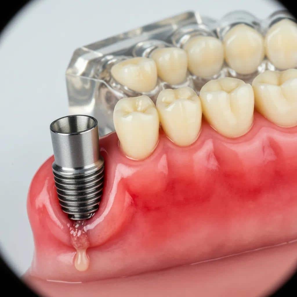
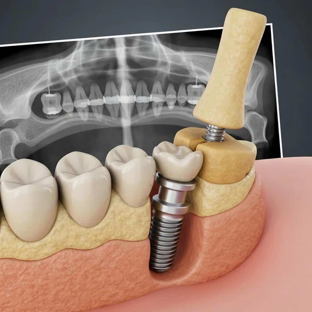
Dental implants replace missing teeth by anchoring a titanium fixture into the jawbone and restoring chewing function and aesthetics through an abutment and crown. While dental implants have a high overall success rate, understanding possible complications—such as infection, nerve injury, sinus issues, and graft failure—helps patients make informed decisions and take proactive steps to reduce risk. This article explains the most common risks associated with dental implant surgery, the signs of implant failure, and concrete prevention and recovery strategies you can apply before and after surgery. You will find evidence-based descriptions of nerve injury, peri-implantitis, sinus perforation, and bone graft concerns, plus practical pre-surgical checklists, post-op care guidance, sedation options, and clear red flags that require urgent attention. The piece also includes comparison tables that contrast early versus late implant failure, sinus lift versus bone graft risks, and sedation modalities to support informed consent and planning. If you are considering dental implants, this guide will prepare you to recognize symptoms early, optimize health before surgery, and know what to expect from follow-up care.
What Are the Most Common Risks of Dental Implant Surgery?
Dental implant surgery carries both surgical and biological risks that vary by timing and underlying patient factors, from immediate intraoperative complications to late inflammatory conditions. The principal risks include peri-implant infection, nerve injury, sinus membrane perforation, implant failure (lack of osseointegration or late loss), and bleeding or anesthesia-related events; each arises from distinct mechanisms such as bacterial contamination, anatomical proximity, or poor bone quality. Recognizing these risks and their typical timing helps patients and clinicians prioritize preventive measures like imaging, sterile technique, and medical optimization before the procedure. Below is a concise list of the most frequently encountered risks, followed by short definitions and typical early warning signs to aid rapid recognition and response. Early awareness improves outcomes because many complications are more readily treated when identified promptly.
Peri-implantitis and implant infection: Bacterial inflammation around the implant often causing swelling and bleeding.
Nerve damage: Injury to the inferior alveolar or mental nerve producing numbness or altered sensation.
Sinus perforation: Breach of the sinus membrane during upper jaw implant placement, causing sinus symptoms.
Implant failure: Failed osseointegration or progressive bone loss leading to mobility.
Bleeding and anesthesia reactions: Excessive intraoperative bleeding or adverse responses to sedation/anesthesia.
These risks are distinct but interrelated; appreciating their mechanisms leads to targeted preventive steps that we discuss next and through the rest of the article.
What Causes Dental Implant Infections and How Can You Recognize Them?
Dental implant infections arise when bacteria colonize the peri-implant sulcus or graft site, and factors such as poor oral hygiene, pre-existing periodontal disease, smoking, or systemic conditions increase susceptibility. Infection can present early, within days to weeks of surgery, as an acute surgical site infection with pain, swelling, erythema, and possible purulent discharge, or later as peri-implantitis with progressive bone loss and pocket formation. Diagnosis typically combines clinical signs—tenderness, suppuration, and bleeding on probing—with radiographic evidence of bone loss and, when necessary, microbiological sampling to guide therapy. Immediate patient actions include improving localized cleansing, contacting the clinician for evaluation, and following prescribed antimicrobial or debridement protocols; delayed treatment increases the chance of implant loss. Understanding these causes and recognition patterns supports rapid clinical triage and reduces the likelihood of late-stage complications.
How Does Nerve Damage Occur During Dental Implant Surgery?
Nerve damage during implant placement usually results from direct trauma, compression by the implant body, or excessive drill depth near anatomical sensory nerves, most commonly the inferior alveolar nerve or mental nerve in the lower jaw. Poor three-dimensional planning, inadequate imaging, and failure to account for anatomic variation increase risk; conversely, preoperative CBCT imaging and careful depth control reduce the likelihood of nerve proximity errors. Clinically, nerve injury manifests as immediate or delayed numbness, tingling, burning sensations, altered taste, or dysesthesia in the lip, chin, or tongue distribution; persistent paresthesia beyond expected temporary anesthesia warrants prompt evaluation. Management options range from observation for partial sensory recovery to medication, referral for neurosensory testing, or surgical intervention for decompression or implant removal if a compressive etiology is identified. Early detection and timely referral improve prognosis, which is why precise planning and patient counseling are essential before implant surgery.
Inferior Alveolar Nerve Injury in Dental Implant Placement: Diagnosis and Treatment
The purpose of present article was to review aetiological factors, mechanism, clinical symptoms, and diagnostic methods as well as to create treatment guidelines for the management of inferior alveolar nerve injury during dental implant placement.
Injury of the inferior alveolar nerve during implant placement: a literature review, G Juodzbalys, 2011
What Are the Signs and Symptoms of Dental Implant Failure?
Implant failure refers to loss of the implant’s structural or biological integration with surrounding tissues, presenting as mobility, persistent pain, or progressive bone loss that compromises function and aesthetics. Failure can be divided conceptually into early failure (failure to osseointegrate within months of placement) and late failure (loss of a previously integrated implant due to infection, overload, or systemic factors), each with distinct causes and timelines. Clinicians use a combination of clinical testing—mobility checks, probing, and soft-tissue assessment—and radiographs to confirm bone loss or peri-implant defects, which guide decisions about conservative management versus revision. The table below compares early and late failure across causes, timing, symptoms, typical treatment, and prevention strategies to help patients understand differences and what to watch for after surgery. Recognizing these differences helps prioritize timely intervention and may preserve surrounding bone and future implant options.
Introductory comparison table describing early vs late implant failure:
Persistent pain, lack of stability, delayed healing
Late Failure
Peri-implantitis, overloading, systemic disease
Months to years after restoration
Mobility, progressive bone loss, recurrent infection
Peri-implantitis (linked)
Bacterial biofilm, poor hygiene, smoking
Can be early or late
Bleeding on probing, suppuration, increasing pocket depth
How to Identify Early vs. Late Dental Implant Failure
Early implant failure stems from unsuccessful osseointegration due to factors such as surgical contamination, micromovement during healing, inadequate primary stability, or compromised bone quality, and it typically appears within the first few months after placement. Signs of early failure include persistent postoperative pain, mobility or lack of stability when prosthetic loads are applied, and delayed soft-tissue healing that does not follow the expected recovery trajectory. In contrast, late failure occurs after an implant has initially integrated and is often caused by peri-implantitis, mechanical overload, or systemic health deterioration; clinical cues include gradual loss of bone on radiographs, increasing pocket depth, and episodic infections. Clinicians determine the appropriate response through clinical examination and imaging: early failure may require implant removal and re-grafting, while late failure management commonly focuses on infection control, debridement, or prosthetic adjustment to reduce overload. Timely identification differentiates salvageable situations from those needing more extensive reconstructive work.
Inferior Alveolar Nerve Injury After Dental Implants: A Systematic Review of Diagnosis and Treatment Outcomes
The purpose of this article is to systematically review diagnostic procedures and risk factors associated with inferior alveolar nerve injury following implant placement, to identify the time interval between inferior alveolar nerve injury and its diagnosis after surgical dental implant placement and compare between outcomes of early and delayed diagnosis and treatment given based on case series recorded throughout a period of 10 years.
Inferior alveolar nerve injuries following implant placement-importance of early diagnosis and treatment: a systematic review, G Juodzbalys, 2014
What Long-Term Problems Can Arise from Failed Dental Implants?
Failed implants can produce several long-term oral health problems, including progressive alveolar bone loss that complicates future implant placement, damage to adjacent teeth, altered occlusion, and chronic soft-tissue infections that reduce overall oral comfort and function. Prosthetic complications—such as loose abutments, fractured restorations, or misfit crowns—often accompany biological failure and may require complex corrective work, including removal and replacement of components or full prosthetic redesign. Chronic peri-implant infection can lead to sinus involvement in the maxilla or spread of infection in vulnerable patients, requiring additional surgical interventions and extended antimicrobial therapy. Treatment pathways range from conservative soft-tissue therapy and localized debridement to surgical grafting, staged re-implantation, or alternative prosthetic solutions depending on bone volume and patient health. Prevention and prompt management of early signs significantly reduce the probability of these long-term sequelae and preserve future treatment options.
How Can You Prevent Complications Before and After Dental Implant Surgery?
Prevention of implant complications combines rigorous pre-surgical assessment, patient optimization (medical and lifestyle), precise surgical technique, and consistent post-operative care designed to support osseointegration and soft-tissue health. A structured approach starts with a comprehensive medical and dental history, high-resolution imaging to evaluate bone volume and anatomical risks, and periodontal treatment when active gum disease is present; optimization of systemic conditions such as diabetes and smoking cessation materially improves healing outcomes. Surgical planning that incorporates guide-based placement, sterile technique, and minimally traumatic handling of soft tissue and bone reduces contamination risk and mechanical stress during healing. Post-operative protocols emphasize pain control, infection prevention, gentle oral hygiene practices, dietary modifications, and scheduled follow-up visits to detect early warning signs; adherence to these steps is a strong predictor of long-term success. Below is a practical, step-by-step preventive checklist to follow before and after implant surgery to minimize complications and promote predictable results.
Complete pre-op medical and dental evaluation: Review systemic health, medications, and periodontal status to identify modifiable risks.
Obtain 3D imaging when indicated: Use CBCT or equivalent imaging to map nerve positions and bone volume for accurate planning.
Optimize systemic and lifestyle factors: Achieve glycemic control if diabetic, stop smoking, and follow medication guidance.
Follow sterile surgical protocols and guided placement: Minimize surgical trauma and bacterial exposure for better osseointegration.
Adhere to post-op care and follow-up schedule: Maintain prescribed oral hygiene, medication adherence, and timely clinical reviews.
Following this checklist lowers infection risk, limits mechanical causes of failure, and supports healthy osseointegration. If you are trying to schedule a free consult to have risks assessed and receive a personalized pre-surgical plan, that is the next sensible step to translate these preventive measures into a tailored treatment pathway.
What Pre-Surgical Assessments Help Minimize Implant Risks?
Effective pre-surgical assessment combines a detailed medical and dental history with targeted imaging and periodontal evaluation to stratify risk and guide implant planning. Medical screening focuses on conditions and medications that affect wound healing or bone metabolism, while dental evaluation assesses remaining tooth support, periodontal stability, and occlusal forces that could impact implant longevity. High-resolution three-dimensional imaging identifies anatomic limitations—nerve canals, sinus anatomy, and bone volume—enabling selection of implant length, angulation, and need for grafting or a sinus lift. A practical pre-op checklist should include documentation of smoking status and glycemic control, review of anticoagulant or bisphosphonate use when present, and stabilization of active oral infections; this reduces avoidable complications. Clear communication between patient and surgical team during assessment sets expectations, informs consent, and allows for tailored strategies to reduce the risk of intraoperative or postoperative adverse events.
What Post-Operative Care Steps Reduce the Risk of Implant Problems?
Post-operative care focuses on protecting the surgical site, controlling infection, managing discomfort, and supporting bone and soft-tissue healing during the critical osseointegration period. Immediate measures include rest, head elevation, application of cold packs to reduce swelling, adherence to prescribed analgesics and antibiotics when indicated, and eating a soft diet while avoiding actions that create negative pressure at the surgical site. Gentle oral hygiene with non-abrasive rinses and careful cleaning around adjacent teeth prevents biofilm accumulation without disrupting clots or sutures; patients should follow clinician instructions for when and how to resume normal brushing and flossing. Scheduled follow-up visits allow clinicians to monitor healing, remove sutures if present, and address early signs of infection or instability; following these appointments and reporting concerns promptly enhances the chance of successful long-term outcomes. Consistent, evidence-based post-op care remains one of the most effective tools patients have to reduce implant complications and preserve oral health.
What Are the Specific Risks Related to Sinus and Bone Grafting Procedures?
Sinus lift and bone graft procedures are common adjuncts to dental implant therapy when native bone volume or height is insufficient, and each procedure carries unique risks that patients should understand before consenting. Sinus-related risks include sinus membrane perforation, graft infection that leads to sinusitis, and postoperative sinus symptoms such as congestion or fluid drainage; bone graft risks include infection, graft exposure, lack of vascularization causing resorption, and donor-site morbidity when autografts are used. Surgical technique, the health of the sinus and oral mucosa, and adequate vascular supply determine the likelihood of graft success; planning with three-dimensional imaging helps identify potential anatomic challenges. Early recognition of symptoms—persistent pain, unusual nasal discharge, or exposed graft material—prompts timely intervention, ranging from conservative antibiotics and observation to surgical revision or graft removal. The table below compares sinus lift and bone graft procedures across indications, common complications, signs to watch for, mitigation strategies, and salvage options to clarify trade-offs and expected post-op courses.
Introductory comparison table for sinus lift versus bone graft risks:
Procedure
Indication
Common Complications
Signs to Monitor
Sinus Lift
Insufficient posterior maxillary height for implants
Improved symptoms after intervention indicate salvage success
How Does Sinus Perforation Happen During Implant Placement?
Sinus membrane perforation occurs most often when implant osteotomies or graft placements extend too close to or through the maxillary sinus floor, particularly when residual bone height is limited and the clinician underestimates membrane position. Risk factors include thin or fragile Schneiderian membrane, aggressive instrumentation, lack of adequate imaging, or repeating instrumentation in the same area during grafting. Immediate signs of perforation can range from an audible change during the procedure to postoperative sinus symptoms such as nasal regurgitation of oral fluids, unusual nasal discharge, or persistent sinus congestion and pain. Management strategies depend on perforation size and condition: small perforations are often repaired with collagen membranes and careful postoperative monitoring, whereas larger tears or established sinus infection may require postponement of implant placement and ENT consultation. Preventive measures like pre-op CBCT imaging, atraumatic elevation techniques, and having contingency plans for membrane repair reduce the incidence and improve outcomes when perforation occurs.
What Are the Risks and Signs of Bone Graft Failure?
Bone graft failure arises from inadequate vascularization, contamination at the graft site, mechanical instability, or patient factors such as smoking and uncontrolled systemic disease that impede healing. Clinically, failing grafts present with prolonged pain, localized swelling, graft exposure through the mucosa, and lack of expected radiographic integration over follow-up visits; soft-tissue breakdown over the graft is a particular warning sign that infection or resorption is occurring. Management typically begins conservatively with improved oral hygiene, antimicrobial therapy when indicated, and removal of exposed nonviable material; persistent failure may necessitate graft removal, re-grafting with augmented technique, or alternative prosthetic planning. Preventive strategies include selecting graft materials and techniques matched to defect size, ensuring primary wound closure, minimizing contamination, and optimizing patient health before surgery. Early diagnosis and timely intervention increase the chance of salvage and reduce the cascade of complications that can complicate later implant attempts.
How Does Patient Health Affect the Risks of Dental Implant Surgery?
Systemic health profoundly influences implant outcomes because wound healing, bone remodeling, and immune response determine the success of osseointegration and the ability to resist infection. Conditions such as diabetes, smoking-related vascular compromise, osteoporosis or low bone density, and certain medications (for example, antiresorptives) alter bone metabolism and soft-tissue healing, increasing risk for both early and late implant complications. A thorough optimization plan that addresses glycemic control, smoking cessation, medication review, and bone quality assessment helps clinicians tailor the timing and type of implant therapy to minimize risk. Clinics offering pre-surgical assessments and individualized plans can help patients identify modifiable risks and select appropriate adjunctive measures such as bone grafting or staged surgery; financing flexibility and sedation options further support access and comfort during necessary preparatory steps. Assessing and improving patient health before surgery reduces complication rates and improves long-term implant survival by aligning biological readiness with surgical choices.
When discussing patient optimization, the clinic can play a valuable role by offering pre-surgical assessments and tailored plans that consider medical history, imaging, and lifestyle factors. Dentist In Mansfield– Dentures and Dental Implants provides pre-surgical assessment pathways, personalized treatment planning, and options for comfort through sedation dentistry; the practice also offers financing options and an Affordable Discount Plan to help patients proceed with necessary preparatory care.
How Do Diabetes and Smoking Increase Implant Complications?
Diabetes impairs wound healing through hyperglycemia-driven changes in immune cell function and microvascular perfusion, which increases susceptibility to infection and delays bone remodeling necessary for osseointegration. Elevated blood glucose correlates with higher rates of peri-implantitis and implant failure when glycemic control is poor; clinicians commonly recommend achieving stable glycemic markers before elective implant surgery to reduce this risk. Smoking introduces systemic and local vasoconstriction, impairs oxygen delivery, and promotes biofilm formation, all of which hinder mucosal healing and bone regeneration and raise failure rates compared with non-smokers. Practical patient optimization steps include achieving diabetes control targets as advised by a treating physician, engaging in smoking cessation programs well before surgery, and following clinician guidance on medication and lifestyle adjustments to support healing. Addressing these modifiable factors before implant placement materially reduces the probability of both early and late complications.
Why Is Bone Density Important for Implant Success?
Bone density and quality determine primary stability and the long-term mechanical environment required for osseointegration, making them central considerations in implant selection and timing. Low bone density or deficient ridge dimensions may necessitate augmentation—such as bone grafting or a sinus lift—shorter or wider-diameter implants, or staged surgical approaches to allow sufficient volume and vascularization for integration. Assessment methods like clinical evaluation and three-dimensional imaging identify areas of low density and help guide implant type, length, and the need for grafting; this information also informs patient counseling about prognosis and timeline. When bone is insufficient, augmentation protocols can restore volume, but these add complexity and potential graft-related risks that require careful planning and follow-up. Prioritizing bone health through assessment and appropriate augmentation options improves stability, reduces micromotion at the implant interface, and increases the likelihood of long-term success.
How Can Sedation Dentistry Help Reduce Anxiety and Surgical Risks?
Sedation dentistry provides graded options to reduce patient anxiety, limit movement during procedures, and create stable surgical conditions that can lower the risk of technical errors and intraoperative complications. Different sedation levels range from minimal anxiolysis with local anesthesia to moderate inhalation or oral sedation and monitored intravenous sedation for deeper anxiolysis; each option balances comfort and patient responsiveness against monitoring requirements and recovery time. Sedation reduces physiologic stress responses—such as tachycardia and hypertension from anxiety—that can complicate bleeding control and patient cooperation, and it enables surgeons to work more efficiently and precisely in anxious or medically complex patients. Safety protocols include pre-sedation screening, monitoring by trained staff, and adherence to recovery guidelines to minimize sedation-related events. The comparison table below outlines common sedation modalities, expected depth, typical uses for implant surgery, and safety considerations to help patients and clinicians match the right approach to individual needs.
Introductory sedation comparison table:
Sedation Option
Level of Sedation
Typical Use Case
Safety Notes
Local-only
Minimal (no systemic sedation)
Simple implant placements in calm patients
Routine monitoring; fastest recovery
Nitrous oxide
Minimal-moderate
Anxious patients needing short procedures
Reversible, minimal systemic effects
Oral sedation
Moderate
Patients with moderate anxiety or longer procedures
Variable absorption; requires monitoring
IV sedation
Moderate-deep
Complex surgery or very anxious patients
Requires trained staff and close monitoring
What Sedation Options Are Available for Dental Implant Surgery?
Common sedation options include local anesthesia for pain control, nitrous oxide for mild anxiolysis, oral sedatives for moderate relaxation, and intravenous (IV) sedation for deeper anxiolysis and amnesia during more complex or longer procedures. Local anesthesia blocks sensory input at the surgical site and remains the foundation of pain control for all implant procedures, while nitrous oxide offers quick onset and reversible calming effects suited for shorter visits or needle-phobic patients. Oral sedation provides longer-lasting anxiolysis but has variable onset and metabolism among patients and typically requires escort home after the procedure; IV sedation affords precise titration and deeper sedation levels with rapid adjustment and reliable intraoperative comfort. Safety considerations include pre-sedation medical screening, staff trained in airway management, and post-operative monitoring until patients meet discharge criteria. Selecting a modality is a shared decision between patient and clinician, balancing anxiety level, medical history, procedure complexity, and recovery considerations.
How Does Sedation Improve Patient Experience and Safety?
Sedation improves patient experience by reducing anxiety, minimizing intraoperative movement, and creating a calm operative environment that allows the clinician to focus on precise technique rather than patient comfort. Physiologically, reduced anxiety lowers sympathetic responses such as elevated heart rate and blood pressure, which can otherwise increase bleeding and complicate delicate surgical maneuvers; a stable physiologic state supports more predictable hemostasis and reduced operative stress. Sedation also reduces the memory of the procedure for patients who prefer lower recall, improving overall satisfaction and lowering future procedural avoidance. Safety is maintained through pre-sedation evaluation, appropriate selection of sedation level based on medical history, continuous monitoring during the procedure, and recovery protocols to ensure patients are stable before discharge. When combined with careful planning and surgical technique, sedation is a valuable adjunct that reduces both perceived and clinical risk during dental implant surgery.
What Should You Do If You Experience Complications After Dental Implant Surgery?
If you suspect a complication after implant surgery, prompt action and clear communication with your dental team increase the chance of a successful resolution and reduce long-term damage. Immediate triage prioritizes life- or limb-threatening symptoms such as uncontrolled bleeding, severe spreading swelling, or airway compromise that require emergency care; other urgent signs include fever, severe or worsening pain not relieved by prescribed medication, persistent numbness beyond expected anesthesia, or purulent drainage from the site. For less acute but concerning symptoms—increasing redness, mild drainage, or loosening of the prosthesis—contact your dental provider for an urgent consultation; clinicians will typically assess, possibly prescribe antibiotics, and schedule in-office interventions like debridement or suture revision. A structured approach to reporting symptoms (describe onset, severity, any systemic signs, and provide photos if possible) expedites triage and helps the team plan the next steps. Below is a prioritized action checklist to guide patients on when to seek immediate emergency care versus contacting their dental office for urgent follow-up.
Emergency care now: Uncontrolled bleeding, difficulty breathing, or rapidly spreading facial swelling.
Urgent contact same day: High fever, severe pain not controlled by meds, or signs of spreading infection.
Call for prompt consult: New or worsening numbness, persistent drainage, increasing mobility of implant.
Monitor and document: Take photos, note symptom onset and progression, and follow clinician instructions for medication and wound care.
When to Contact Your Dentist for Possible Implant Problems?
Contact your dental provider promptly if you observe red flags such as uncontrolled bleeding, fever, rapidly increasing swelling, severe unrelieved pain, or new sensory changes like numbness or tingling that persist beyond the expected anesthetic period. For non-life-threatening but concerning signs—increasing redness, persistent discharge, loosening of the restoration, or delayed wound closure—call for an urgent appointment so clinicians can evaluate and intervene with conservative measures such as antibiotics, cleaning, or prosthetic adjustments. When calling, provide a clear description of symptoms, onset timing, any systemic signs (fever, malaise), photos if possible, and recent medication use; this information expedites triage and helps the team prioritize care. Expect the clinic to advise immediate steps you can take at home to reduce risk (e.g., apply pressure for bleeding, avoid rinsing vigorously) and to schedule an evaluation to determine whether in-office treatment or emergency referral is needed. Acting early improves the likelihood that conservative measures will be effective and reduces the need for more invasive interventions.
What Treatment Options Are Available for Managing Implant Complications?
Treatment options depend on the specific complication but generally range from conservative medical management to surgical revision or implant removal when necessary to protect surrounding structures and restore oral health. For infections and early peri-implant inflammation, clinicians commonly use targeted antibiotics, improved local hygiene measures, and mechanical debridement to remove biofilm; adjunctive antiseptic rinses and monitoring support resolution. For mechanical issues such as implant mobility or malposition, options include prosthetic modification, splinting, removal of the failing implant, and staged re-implantation often combined with bone grafting if necessary. Nerve injuries are managed according to severity: observation and medical therapy for mild neuropraxia and referral for surgical intervention if progressive or persistent compression is suspected. Sinus perforations or graft failures may require membrane repair, antibiotic therapy, staged re-grafting, or collaboration with ENT specialists to restore sinus health before further implant work. Timely, complication-specific interventions yield the best prognoses and minimize long-term sequelae.
Why Choose Dentist In Mansfield for Safe and Affordable Dental Implant Surgery?
Dentist In Mansfield- Dentures and Dental Implants focuses on patient-centered implant care that blends education, comfort, and structured pre-surgical assessment to reduce risks and improve outcomes. The practice emphasizes a spa-like, education-based environment with an experienced dental team led by Dr. Mike Tri Pham and Dr. Elvis Le, offering comfort and sedation dentistry options to help anxious or medically complex patients undergo implant procedures with appropriate monitoring and support. The clinic provides practical access solutions including a free dental implant consult to assess individual risk, review imaging, and present personalized treatment pathways so patients can make informed choices. Financing options are available, including flexible payment plans, low-interest installment arrangements, and an Affordable Discount Plan that can provide meaningful savings, making implant therapy more accessible without compromising on safety or quality. For patients seeking dental implants, this combination of pre-surgical assessment, sedation comfort, and financial flexibility supports safe, affordable care tailored to individual needs.
How Does Our Experienced Team Minimize Implant Surgery Risks?
The team approach at Dentist In Mansfield prioritizes thorough pre-op evaluation, clear communication, and protocols designed to reduce infection, nerve injury, and graft-related complications. Clinicians conduct pre-surgical assessments that include medical and dental histories, imaging review, and risk stratification to determine the need for adjunctive procedures such as grafting or staged placement; this planning reduces intraoperative surprises and guides the selection of sedation and monitoring. Sedation dentistry and comfort-focused techniques are available to manage anxiety and minimize patient movement during placement, which can reduce technical errors and improve surgical conditions. Postoperative follow-up protocols emphasize early monitoring for infection or healing problems and scheduling timely interventions when needed, reinforcing continuity of care. These process-oriented safeguards help align surgical technique with patient-specific risks to support predictable outcomes.
What Financing and Free Consultation Options Are Available?
For patients considering implant treatment, Dentist In Mansfield offers a complimentary dental implant consult that evaluates candidacy, explains risk-reduction strategies, and outlines personalized next steps to move forward with confidence. The practice also provides a range of financing options, including flexible financing plans and low-interest installment arrangements, as well as an Affordable Discount Plan that can reduce overall treatment costs by 30–60 percent for eligible services. These financial tools are intended to make necessary preparatory care, grafting, and implant placement more attainable while preserving high standards for safety and patient comfort. To determine eligibility and discuss tailored financing alongside a risk assessment, scheduling a free consult is the recommended first step for anyone planning dental implant surgery.